|
The most of time in sciences, reasearchers publish article or review in sciences journal as for example Nature, Sciences or more specific journal as Biomaterials Science, Cells Biology etc … (Fig 1). However, it is not the only possibitility. In certain case, it is more interessant to register a patent. What are the differences between a patent and an article? Why it can be interessant to register a patent?
Figure 1: Example of sciences journal [1,2,3]
Publications in sciences are very important because it allows for researchers to show their work and to leave a proof of their work inside the science community. However, to publish an article, it takes time and costs some money. It is necessary to manage to find a journal which agrees to publish our article. Then, the article is reviewed by other scientists who are going to suggest modifications. Finally, when the publisher of the journal is in agreement with the contents and the shaping of the article, this one can be finally published. The process of publication of an article sets several months and can even last more than year in certain cases. Besides long necessary deadline to publish an article, it is necessary to add costs which are raised. These costs vary according to the raputation of the journal, the size of the article, etc.... The publication of an article is thus a process which is long and expensive, it is thus necessary to think before deciding to publish an article. During the register of a patent, we find the same problems. Actually, to apply for a patent is a process which is long and which costs some money. However, a patent is very different with a scientific article at the level of the contents. A patent is a way to protect an invention. To apply for a patent, it is necessary to have a new technique answering a problem. It is very important that the technique is new because otherwise the patent cannot be accepted. During the register of a patent, thus we are going to make a description to explain our new technique, how it works, which are the innovations which we bring and what is the use of this new technique. We are also going to make claiming which allow to explain what are the conditions of use of the technique. We also have knowledges which are sorts of "advice" on the use of the invention. Thus the patent allows to protect an invention. Indeed, when it is register, if somebody wants to use your technique, he will have to have the authorization to use it freely either pay a sum to the inventor of the technique. To apply for a patent, it is necessary to address to a office specialized in the register of patents. We have to show them that our technique is new. Then, it realizes a study of the file then gives their decision onto the register of the patent. It is necessary to supply many results to prove that the invented technique is new and works correctly. If the patent is accepted, it is the office which realizes the writing of the patent in a special language with a characteristic shaping of the writing of patents. It is possible to apply for a national, European or international patent. Naturally, the costs are not the same according to the level of the patent which we wish to deposit. The register of a patent is thus the cause of many problems of confidentiality. Indeed, it is the first one to have applied for the patent which benefits from everything the rights. It is thus necessary to be sure that the realized work remains confidential to avoid that other people apply for a patent before your. We also find this type of problems with scientific articles. The researchers have to watch the confidentiality of their results to avoid that another team publishes before them. You will thus have understood it, a scientific article allows to show the advanced of its researches for a scientist and to leave a proof of its work in the scientific community whereas a patent is a way to protect the invention of a new technique and we can ask some money in the exchange of the use of our invented technique. The redaction of a scientific article and a patent are completely different and are especially realized in different purposes. However, in both cases, we are faced with processes which are long and expensive and thus ask to be reflected well before being begun. We will have to pay also attention on the confidentiality of the data. Thank you for you reading! See you soon :) References: [1]http://umdrightnow.umd.edu/news/starburst-wind-keeps-galaxies-thin [2]http://pubs.rsc.org/en/journals/print?sercode=bm&issueid=bm004001&journalname=biomaterials%20science&isarchive=false&issnprint=2047-4830&pagenumber=1&tabname=issues [3]http://biomachina.org/research/projects/actin/
3 Comments
For my internship which concerns the bone regeneration, I had the opportunity to be able to discover the cell culture. Some of you think maybe that the cell culture in microbiology is similar to the other types of cell cultures but they exist differences. We are going to begin by approaching the microbiology with a fast reminder of the characteristics of this domain because you already know it a lot about this subject then we will speak of other types of cell cultures and their differences with the microbiology. Microbiology The microbiology is the domain which studies microorganisms. We study bacteria, fungis, algaes or viruses. In this domain, we thus study as well procaryotes organisms as eucaryotes organisms (Fig 1). [1]
Figure 1: Bacteria [2] and Yeast [3] representation Very often in microbiology, we study bacteria and yeasts because they are model organisms and easy to study thanks to different advantages. For example, their genomes is totally sequenced, their cycles of division is fast, they are easy to maintain in culture, etc. … Indeed, in microbiology, it is an advantage to study microorganisms because it simplifies the studies. In general, conditions to study microorganisms are not very complicated to be gathered. It is enough to realize rich media as LB media (lysogeny broth or media Luria-Bertani) which is a media used for the bacterial culture. [4] We can also quote the YPD media (Yeast extract Peptone Dextrose) which is a rich media for yeast culture. [5] However, we do not use only rich media in microbiology, we can also use poor media in order to create certains mutation in the studied colonies. We can make the cultures in liquid media in flasks or in solid media with petridish. Microorganisms are often also easy to study because they can possess various resistances on the conditions in which they live. There are for example bacteria living in very acid environment either in high temperatures. It is necessary to maintain regulary the colonies which we cultivate as by changing the media of culture but it is fast and does not necessarily ask to be made every day. The regulations for the culture of microorganisms depend in particular if we use it organisms modified genetically and if they are pathogenic or not. According to the degree of danger, it is going to be necessary to fit out the room of culture for example by putting an airlock of access, an independent system of aeration, etc… The material used in microbiology is petridishes which serve to cultivate microorganisms as bacteria, flasks are use also, biological hood (PSM), oeses, etc... (Fig 2). We always work in sterile conditions with a PSM or on a flame to avoid the contaminations of cultures.
Figure 2: Petridishes [6] and oeses [7] The microbiology is a vast domain allowing many applications. We can very well used the microbiology to study the present mechanisms in microorganisms, we can also use microorganisms as bacteria to use them in genetic engineering for applications in biotechnologies. We can also study the viruses for applications in biomedical research. The subjects and thus the applications are really varied and we can use mircroorganisms in many different case of studies. Other type of cells culture The cell culture includes all the techniques allowing to cultivate in vitro cells, that is why we find inside the study of microorganisms as bacteria or yeasts. However, the cellular cuture also includes the culture of animal or human cells. [8] There are many types of cells. We can classify them according to their origin and according to the specificities. To start, we distinguish the animal cells of human cells. We are also going to separate according to if it is stem cells, primary cells or immortal lineages. A stem cell is a cell capable of differing in any type of tissues as muscular, osseous, neuronal or cardiac tissues. We can obtain stem cells by taking them directly from foeti, from blood of the umbilical cord or from embryos. We can also obtain stem cells by realizing genetic manipulations on adults cells which consist in undifferenciate adults cells to make become again stem cells and this cells are called induced pluripotent stem cells (iPSCs). There are several ethical problems about the use of stem cells in particular on the way of extracting them. [9] A primary cell is cell that we extracted directly on an organism. This type of cells have the particularity to undergo the senescence and thus die after a certain time. It is also necessary to take many precautions at the time of their extractions to avoid the contaminations by microorganisms. [10] The immortal cells are lineages which cannot age and thus not die in time. They can be obtained from various manners. For example, a lineage of cells can be immortal in the case of cancer cells, if they are infected by a virus making them immortal (ex: herpes virus, SV 40) or by genetic modifications. [11] We will use a type of cells according to the study which we realize. For example, in tissular engineering, we use very often stem cells because they are going to be interesting for the study on tissues regeneration. On the contrary, for other studies, for example it will be interesting to use immortal lineages because we can cultivate them infinitely without they die by apoptose. In the same way as the culture of microorganisms, we also use rich and poor media to cultivate the other types of cells. However, cultures are realized in liquid media in flask or in petridishes. The animal and human cells ask a particular maintenance. Indeed, it is necessary to begin by defrosting them then realizing passages. It corresponds simply to leave our cells in cultures to let them multiply. When we make a passage of our cells, it is made in a certain percentage of confluence according to the study which we realize. However, we are obliged to pass cells before having reached 100% of confluence to have better results. The number and the frequency of the passages depends on the density of cells which is present in the culture. [12] At the level of the material, it is a little bit different that for the culture of microorganisms on certain points. We also work under a PSM in sterile condition, we also use petridishes and incubators. On the other hand, we use special flasks or 96, 48, 24 or 6 well plates for the culture (Fig 3). 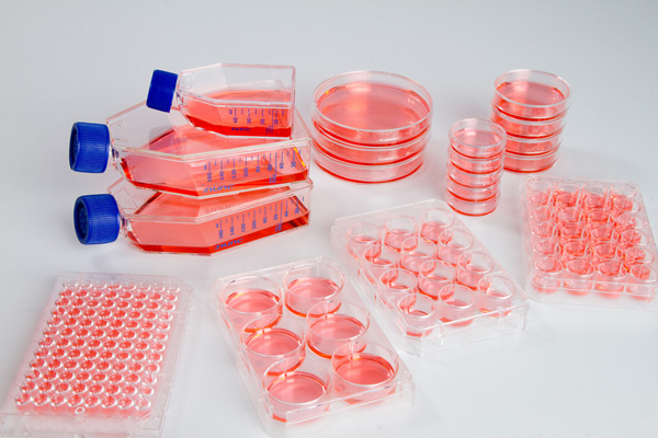 Figure 3: Materials for cell culture [13]
The study of the animal and human cells show themselves very complex. Indeed, cells are sensitive and their resistance are very limited. It is always necessary to maintain them and to cultivate them in good conditions. Furthermore, their studies is complex because the size of their genome and the number of mechanisms which they possess is more raised than those of the microorganimes. At the level of the regulations, it is as the study of microorganisms. Certain rules must be respected and according to the type of cells whom we use, the room of culture must be adapted. It changes in particular if we cultivate stem cells or if we cultivate cells modified genetically. The applications with the human or animal cells and that they are primary, immortal cells or origins are really very vast. Indeed, we can realize many theoretical studies to try to understand many mechanisms but we can also make studies very applied for example to treat diseases as genetic diseases or cancers. Conclusion To conclude, the animal or human cell culture and microorganisms culture have common points but also differences. The principle is the same, we use rich or poor media in liquid or solid condition to cultivate cells. We have to pay attention in both cases on the contaminations. The used material is often the same except for some elements. In both cases, we have to make a strict regulations to cultivate cells. The main differences are situated at the level of the maintenance of the cultures, the complexity to study the organism. Indeed, the animal and human cells are more complex to study from part their biological properties as the size of their genomes but also by their sensibility and their resistances which is lower than microorganimes. Finally, that is with microorganisms as bacteria and yeasts or animal and human cells, their studies allow to answer numerous questions and applications on different subjects. Thank you for you reading and don't hesitate to make comments! See you! References [1] https://fr.wikipedia.org/wiki/Microbiologie [2] http://www.futura-sciences.com/sante/dossiers/biologie-bacteries-leur-monde-nous-1433/page/4/ [3] https://fr.wikipedia.org/wiki/Saccharomyces_cerevisiae [4] https://fr.wikipedia.org/wiki/LB_%28milieu_de_culture%29 [5] https://en.wikipedia.org/wiki/YEPD [6] http://www.usinenouvelle.com/expo/boites-de-petri-o1616.html [7] http://www.sawyoo.com/post_loop-clip-art_706829/ [8] https://fr.wikipedia.org/wiki/Culture_cellulaire [9] http://www.inserm.fr/dossiers-d-information/cellule-souche [10] https://fr.wikiversity.org/wiki/Culture_cellulaire/Culture_de_lign%C3%A9es [11] https://fr.wikiversity.org/wiki/Culture_cellulaire/Culture_primaire [12] https://fr.wikipedia.org/wiki/Passage_%28biologie%29 [13] http://www.dutscher.com/frontoffice/product?produitId=0A-30-08 Today, I am going to present you the subject of my internship which is: the regeneration of the bone! As you suspect it, I did not present you the domains of biomaterials and tissular engineering previously by chance. Indeed, while combining these two domains we let us can regenerate bones. First of all, the laboratory in which I work is the LMGP (Laboratoire des Matériaux et du Génie Physique) within the team IMBM (Interfaces between Materials and Biological Matter). This laboratory is situated inside the scientific pole of Grenoble (see Fig 1.). 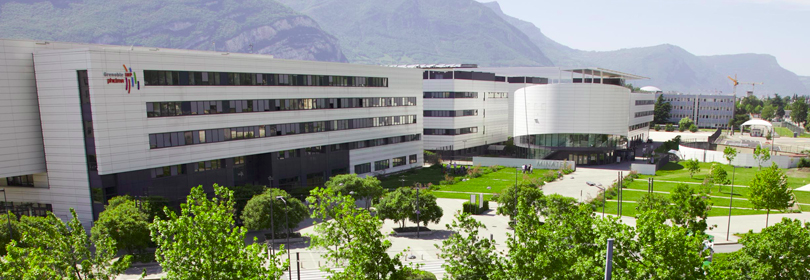 Figure 1: the laboratory LMGP and the scientific pole of Grenoble [1] I am going to begin by explaining to you briefly how we work on the regeneration of the bone and then I would explain you more in detail the used techniques. My subject in some lines Then how it is possible to regenerate bones? It is possible with the regenerative medicine! In fact for it, we are going to combine techniques connected in biomaterials and to the tissular engineering. Indeed, the principle consists in forming films of polymers about implants which we load then with a protein which is a growth factor. It is going to allow the cells which are going to adhere on the implant to be in touch with the growth factor which is going to lead the differentiation of cells in osteoblasts that is more simply osseous cells. Here is a short video in French during a conference TED of my supervisor of internship explaining with vulgarisation our project [2]: The formation of the films of polymers To start, a polymer is a very long chemical molecule formed by the repetition of a motive which we call monomer. The films of polymers which we realize are called PEM (Polyelectrolyte Multilayer films). The polymers which we use are thus polyelectrolytes, they have a charge. We use poly-L-Lysine (PLL) as polycation (charged +) and the polyanion (charged -) is the Hyaluronic Acid (HA) (see Fig 2.). It is thanks to the interaction between these charges that we are going to be able to form our films of polymers. The PLL and the HA are bought under forms of powders then are dissolved in a buffer solution in a precise pH.
Figure 2: Chemical formula of poly-L-Lysine (PLL) [3] and Hyaluronic Acid (HA) [4] The technique which we use to form our films of polyelectrolytes is called LbL for Layers By Layers technique deposition (see Fig 3.). As his name indicates it, we form films by forming successive layers of polymers. The films which we realize are films of PLL/HA. We put a layer of polycation which is the PLL then the layer of polyanion which is the HA and so on to form our films. In every stage of deposition of a layer, we realize rinsings so that films can form correctly. We work with films of 12 or 24 bilayers. We can make films on glasses slides or directly on implants by using various techniques as the dipping which consists in dipping samples successively into tubs containing the various solutions or still by using a spray to apply the solutions. Every technique possesses advantages and inconveniences which play a role on the properties of the film, the cost or the precision. We can also realize various stages of treatment of the film after its formation to modify its properties as for example by realizing a chemical treatment to change its rigidity. We analyze our films in the confocal microscope by marking our film with various fluorescent molecules. So, we can measure the thickness of our film or still quantify its homogeneity. Films can be formed on various types of implants. We can made it on implants in titanium, ceramic or still polymer. 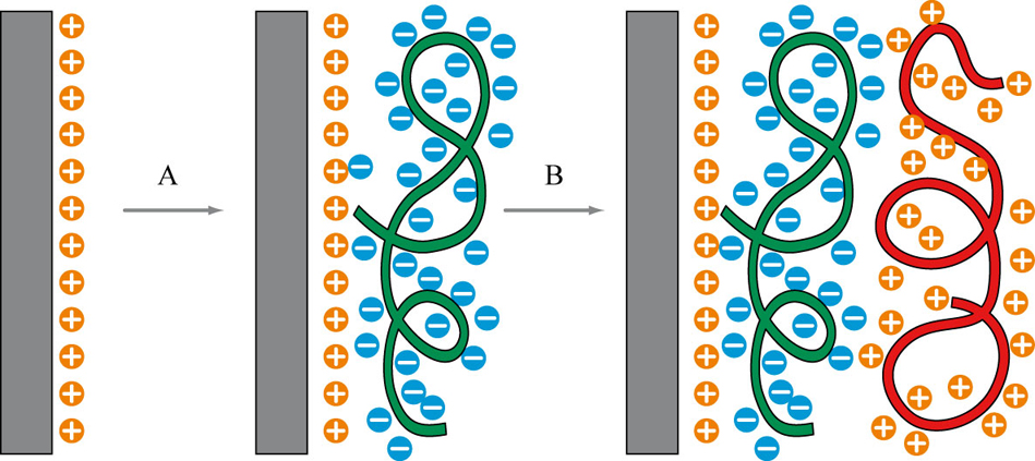 Figure 3: Formation of PEM with the LbL technique deposition [5] Bone Morphogenetic protein 2 (BMP-2) Bones Morphogenetics Proteins are proteins and more exactly growth factors. There are about ten approximately and play in particular an important role in the formation of several types of tissues in the body as the cardiac, muscular, neuronal or still osseous tissues (see Fig4.). For our project, we use the BMP-2 which allows the cells to differ in osteoblasts. Having formed our film of PLL/HA correctly about a glass slide or an implant, we incorporate the BMP-2 into our film.
Figure 4: 3D structure of BMP-2 [6,7]
Finally, the last stage consists in putting cells in cultures on our film for the in vitro studies or otherwise on the implant within a body for the in vivo studies. We can observe the adhesion of our cells with differents types of microscopies as by marking the actin of cells then by analyzing the images in the confocal. We can also verify if our cells differed well by using various tests as enzymatic tests. The majority of the tests which we make are in vitro with cells C2C12 which are cells of mouse which can differ in muscles or in bone. We also use stem cells of mouse. We also already made in vivo tests on rabbits and rats which were a success. We managed to treat fractures in a few weeks while it is normally very difficult. In summary My internship concerns the regenerative medicine and more exactly the regeneration of bones. For that purpose, we form films of polyelectrolytes which we load with a growth factor which allows the cells to differ in osseous cells. We have already obtained from in vitro many interesting results and the first in vivo results are very encouraging. It is thanks to the interdisciplinarity from the team and from the project that we managed to obtain these results by combining the domains of biomaterials and tissular engineering. Thanks to these researches, we have new perspectives for the regenerative medicine because we hope to be able using this technique with other growth factors to be able to regenerate other tissues in the body and also treating cancerous tissues. Thank you for your reading and see you soon! References: [1] http://www.lmgp.grenoble-inp.fr/le-laboratoire/?RH=LMGP_Pr%C3%A9sentation [2] https://www.youtube.com/watch?v=gk7X5gbcz_w&feature=youtu.be [3] http://www.sigmaaldrich.com/catalog/product/sigma/p9155?lang=fr®ion=FR [4] http://www.sigmaaldrich.com/catalog/product/sigma/49775?lang=fr®ion=FR&cm_sp=Insite-_-prodRecCold_xviews-_-prodRecCold10-1 [5] http://eng.thesaurus.rusnano.com/wiki/article1563 [6] https://en.wikipedia.org/wiki/Bone_morphogenetic_protein [7] http://www.rcsb.org/pdb/explore.do?structureId=1REU From the beginning of my internship, I was lucky being able to attend several thesis defenses and also several conferences. Thanks to these moments, I was able to learn many things!
Thus I am going to try to explain why I found this thesis defenses and conferences interesting. During this internship, I was able to attend my first thesis defense. For those of you who do not still know the functioning, the PhD student has to produce his handwritten then to make a defense to obtain its rank of doctor. The defense during approximately 45min then is followed by numerous questions asked by the jury and it can last for a long time (one of the defenses which I attended, the questions during 1h30). The defense takes place in French unless one of the members of the jury does not speak French. The jury is constituted by one or several thesis supervisors of the student and consists of researchers who can be connected directly or indirectly about thesis of the student. The PhD student thus has 45min for summary 3 years of work approximately! It is very interesting to attend a thesis defense all the more if it the subject is connected to your domain of study of your internship. Indeed, it can inspire you for many reasons. We can discover new techniques of manipulations, understand mechanisms which can explain mechanisms connected to our subject, discover new perspectives of applications, etc.... And even if the defense is on a subject which is not necessarily connected in your, it can always give you ideas for the continuation of your internship! When the defense is finished, the jury deliberates to know if they are going to give doctor's title to the student. Then, they give the result and finally a drinks party takes place! This part is also very interesting (and I do not say that because there are champagne and buffet!) because it allows it can discuss with many people that is students in internships, other PhD students or researchers. We can exchange with them, ask numerous questions. It allows us to obtain many advice and even to build up to itself new contacts for future! I was also lucky to attend several conferences from the beginning of my internship. I was able in particular to go to the conference NamiSceb which concerns the subjects of the tissular engineering, the biomaterials and the materials. The conference lasted all day long, I was able to discover students' presentations in theses and researchers showing their projects. I also attended has a session of presentations of posters. This kind of conference is very interesting because once again it allows us to discover numerous different subjects and thus to give us of new ideas for our own projects. Indeed, I was able for example to discover the use of new techniques as the optogenetic, a microscope without lens (the image is a hologram) or still of new applications as the creation of microgels allowing of encapsulate some insulin with the aim of the relarger according to the glycemia of the patient. It would allow to delete insulin pumps! The main conference was given by Nicolas Biais who is a researcher in the field of the mechano-microbiology in the Brooklyn College to The City University of New York. This researcher wanted to show also that it could be very interesting to study procaryotes and not only eucaryotes even for domains as tissular engineering. The studies of this researcher concerned in particular the study of the pilus of bacteria. During this day, I was able to meet and to discuss with many people as well between the conferences as during the session of posters or even during the buffet. It made me discover numerous things and allowed me to have new ideas for my internship. In summary, the thesis defenses and the conferences are moments which allow to discover new things, to give ideas for your projects and to meet new people for your network. I hope to have convinced you to go now to the thesis defenses and to the conferences if you have the opportunity there. Do not hesitate to express your opinion on the subject! Loïc Biomaterials and tissular engineering: a new way for sciencesToday, through this first blog post, I go tried to introduce you and to make you discover the domains of biomaterials and tissular engineering. I am fascinated by these two domains and I have the opportunity to do my internship in a laboratory in the interface of these two domains. I am thus going to explain to you which are the subjects of studies, the issues, the applications and the perspectives for biomaterials and tissular engineering. To start, we are going to define what is one biomaterials and for it we are going to leave the definition given during the conferences of Chester to the United Kingdom in 1986 which is generally admitted by the scientific but also medical community and which indicates a «not alive material, used in a medical device and conceived to interact with biological systems, that it participates in constitution of an equipment with diagnostic aim or with that of a substitute of tissue or organ, or still in that of a device of substitution (or assistance) functional». [1] As you will have understood it, biomaterials are natural or synthetic materials which are going to interact with our body. We can use various types of materials as metals, ceramics or still polymers. We can use them in many applications as the conception of prostheses or artificial organs (see Fig.1) but we can also use it for the production of medicine of which we are going to control the diffusion in the body thanks to the types of used biomaterials or then we can also use biomaterials as support for the tissular engineering.
Figure 1: Example of a prosthesis of ceramic hip [2] and the artificial heart Carmat [3] Biomaterials is an interdisciplinary domain because to study it, we use of the biology for the interactions with the body, the chemistry for the synthesis, the characterization and the reactions of materials but also physics and mathematics for the studies of the applied forces or for the formation and the characteristics of materials. Another important domain is the medicine without whom it would be impossible being able to make accessible biomaterials to the patients. Biomaterials are thus a complex domain to study and which contain many issues. Indeed, we can study the biomaterials of various manners and for different applications. For example, there are many problems in biomaterials as problems of biocompatibility of implants, or still the biodegradability of materials. Actually, it is very complicated to make biomaterials which will have at the same time the properties wanted to treat the patient and which will be harmless as well on the short term as the long term. Furthermore, in these problems are added economic issues because it has to be as much as possible accessible to all. Finally, we can even add ethical issues on questions connected to the use of artificial organs with the fear of having «an immortal Man» or «a superman». Henceforth, let us approach the tissular engineering which corresponds as its name indicates it to the study of tissues. This domain mainly aims at maintaining, at treating or at restoring tissues of the body. We speak very often about regenerative medicine. There are many strategies to study the tissular engineering. For example, we can try to focus its studies on the cell culture and the cellular mechanisms to have a better fundamental knowledge, we can also try to concentrate on applications as to regenerate tissues by combining the use of growth factor during the cell culture or still by using materials with various 3D architectures. There are also many studies on stem cells with the aim of being able to synthesize any type of tissues or treating sick tissues. The tissular engineering is also a very interdisciplinary domain in which is combined the biology and the engineers sciences. This domain is very complex because it is necessary to try to develop high technologies which are on the scale of the micrometer or of the nanometer. Biomaterials and tissular engineering are two domains which can be together used. We can combine them for applications in regenerative medicine. One of the strategies which is more and more developed is the one to use a support in biomaterials on which we are going to put a growth factor which is going to allow for example the cells which come to adhere to its surface to differ in tissues wanted (see Fig.2). However, this kind of study is very complicated because even if we obtain it from good in vitro results, it happens very often that it is much more difficult to set up in vivo. 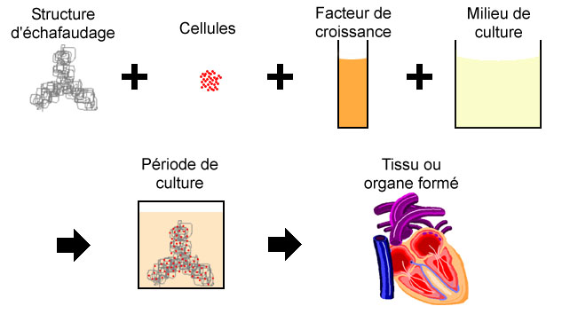 Figure 2: Combination of biomaterials and tissular engineering for the regenerative medicine [4]
Nowadays, biomaterials and tissular engineering are two domains which know a strong development. They are used in many applications as the production of prostheses, artificial organs or still in the regenerative medicine. They also contain important issues which are scientific, economic and ethical. These two domains are really more and more to study and represent a new way for the future of the sciences because the discoveries do not stop increasing and the applications are more and more present and allow every year to treat thousands of people in the world. Now that you know a little more about biomaterials and tissular engineering, I would make you discover my laboratory and my subject of study in the next blog post. Thank you for your reading and see you soon! References: [1] http://www.inserm.fr/thematiques/technologies-pour-la-sante/dossiers-d-information/biomateriaux [2] http://www.chirurgiedelahanche.com/3.aspx?sr=1 [3] http://www.lemonde.fr/sante/article/2016/01/21/mort-du-quatrieme-patient-greffe-du-c-ur-artificiel-de-carmat_4850749_1651302.html [4] http://theses.ulaval.ca/archimede/fichiers/22182/ch02.html |
Search the site...


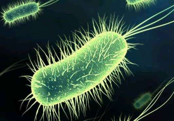
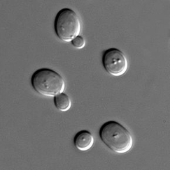

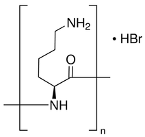
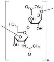
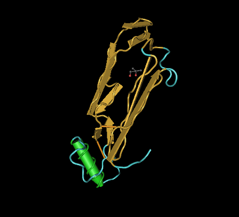
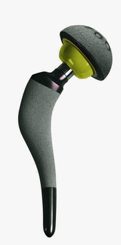
Photo used under Creative Commons from DJANDYW.COM & DJANDYW.TV AKA ANDREW WILLARD
