|
In my laboratory, each Tuesday we have a Lab Meeting of one hours with all people . This week, we spoke of Animals Ethics ! For remember, i realize an internship at Stem cell and Brain Research Institute. Like many laboratory we have animals for the research. So this Lab Meeting was very particular about this sensible subject. In the room of conference there was searchers on stem cell, brain, robotics, administrative people and veterinary, most people had divergence points of view on the subject. This day, the director of research of my lab wanted to speak of new proposition* concerning the large public. Today, we can look horror pictures on animals laboratory on youtube, facebook or other web site. So we formed in our mind a picture of this subject very delicate, a horror vision ! I don’t say this vision is not True, but I want today share with you an other version more adapted for the animals ethics. In Neuroscience we study several subject on animals, each new research protocol which include laboratory animals is presented front of ethic committee which give a validation or not to start the project, in fonction of several criteria ! The criterias can change in function of each project, but keep the same main way. In neuroscience each variation of emotion, or changement of physical aspect on the subjects can have negative effect on the experiment. So if you want a good results, we have to pay attention for our animals. In my team, each animals have a name, games, times and a specially relation with her searcher and a veterinary. So, why inflict a bad treatment on the laboratory animals, when we can have more without that ? * I can't speak of this proposition now ! 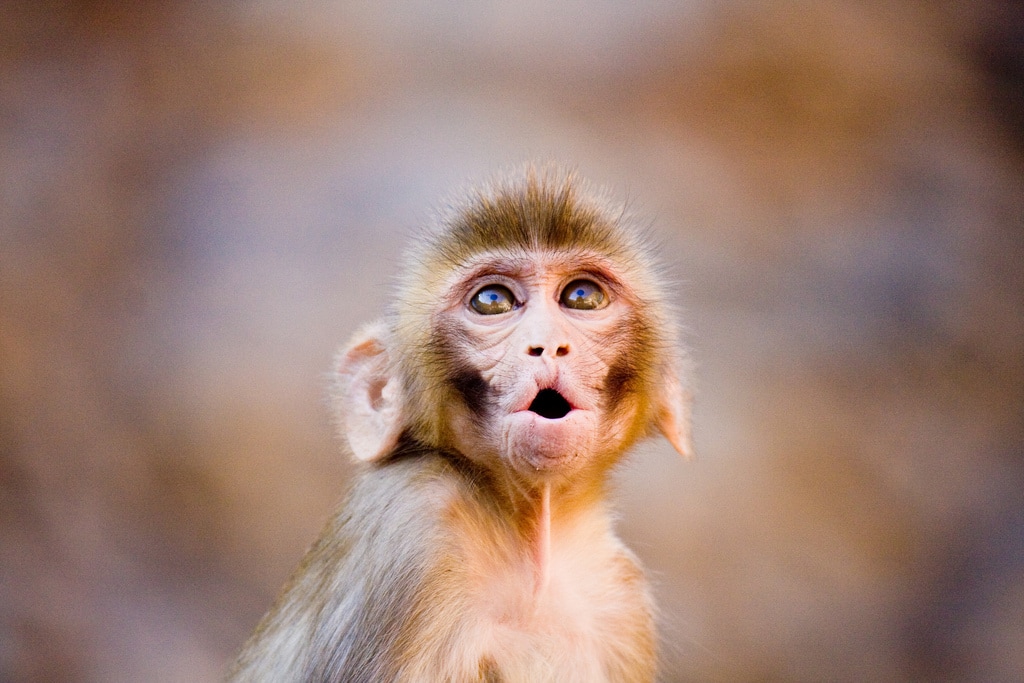 Fig 1 - Rhesus Monkey
Bibliography :
6 Comments
In order to analyse the fMRI datas previously recorded by the team of my laboratory. We use -AFNI- software for analysis and visualization of functional magnetic resonance neuroimages. This software we allow to see in three/four (time) dimensionals the brain. This parameters we allow to see in depth the active area of brain and to target a specific zone, that we want to study. AFNI is an open source software (Linux - Mac - not windows) developed at the Medical College of Wisconsin (1994). Later on the same team developed two other software always in Scientific and Statistical Computer (NIH and NIMH), which can be use in parallel with AFNI. SPM Statistical parametric mapping - SPM is an open source software (mac - linux - windows) which allows to study the difference in brain activity recorded during positron emission tomography. Here, we study fMRI data. When we recuperate the data of fMRI we can not directly analysis the data with AFNI because the data is not on the good format. So we use SPM before AFNI in order to realize a pre-processing on datas. During the pre-processing we parameter the data, in order to have the more beautiful brain mapping. For that we do the better mask (main map) with the aim of to be the more precise. Then, we apply a filter for delete a maximal of noize. In this manner we have great datas to continue our analyse with AFNI. 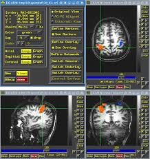 Bibliographie :
Fig 1 - AFNI - AFNI Article - https://www.ncbi.nlm.nih.gov/pubmed/8812068 In my laboratory , we study the connectivity patterns of differents area in the prefrontal cortex (In particular, cingular cortex and Broca area) and their evolution in non-human primates. One of the goal of this study is to obtain a better understanding of the evolution of cognitive capacities (e.g language). My work in this laboratory is to study the IRMf data at resting state obtained on non-human primates. fMRI - Non Human Primate The fMRI (Functional magnetic resonance Imaging) is able to do brain mapping imagery of cerebral activity, this method is based on the detection of the local blood flow and oxygenation changes following neuronal activity (Bold signal, Blood oxygenation level dependent). Indeed, when a region of the brain is active, the demand for oxygen in the neurons increases. There is therefore an increase in blood flow in the active area of the brain.Then since blood oxygenation change according to the level of neural activity these differences can be used to detect brain activity. 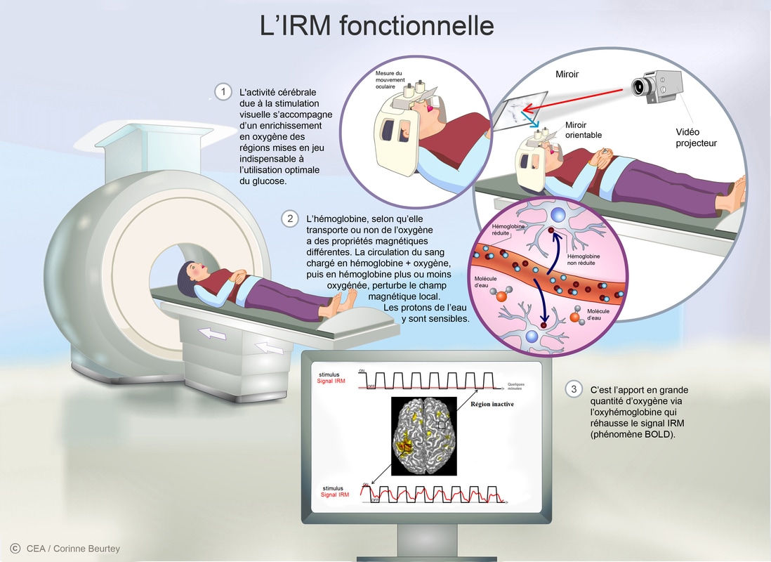 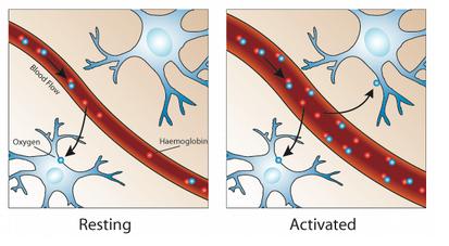 Fig 1 - fRMI Fig 2 - Signal Blood The fMRI can to be used in two conditions with the monkey - Awake or with an anesthesia. We can't do the fMRI if the monkey is asleep because it's dangerous. So we have to do a light anesthesia, because a heavy anesthesia risks to damage the area we want to study. Awake condition : In order to stimulate a specific cerebral area, the subject realizes a cognitive task during its passage in fMRI. This task will allow use to see and record the active cerebral zones . Anesthesia condition - Resting State : The subject doesn’t realize a cognitive task, it's the resting state. This method will allow use to realize an experiment control on the brain. The team realized the resting state method during 1 hour on each of the five monkeys. During my internship, I will concentrate on studying the data of only one monkey named Shirley :) ! Bibliographie : Fig 1 - http://www.cea.fr/comprendre/PublishingImages/Pages/sante-sciences-du-vivant/essentiel-sur-imagerie-medicale/irm-fonctionnelle180413def.jpg Fig 2 - https://amourhainetpe.wordpress.com/la-haine/c-lirm/ There is a lot of answer to this question. However, I can give you one. Once upon a time, in 1905, Campbell described the cytoarchitecture of the human cerebral cortex (1). However, this was a gross description in which he divided only a few general regions. At the same time (1905), Brodmann described the first map of the monkey (cercopithecus) cerebral cortex (2). To ensure a continuum between monkey and human cytoarchitectonic organization of the cerebral cortex, Bromann then described in 1909 the cytoarchitectonic organization of the human cerebral cortex using the same criteria in both species. This map is now the most used in neuroimaging studies, which refer to “Brodmann’s areas”. However, discrepancies remained between the cytoarchitectonic organization of the cerebral cortex in human and monkey. To clarify this question, Dr Petrides, a Canadian researcher, did a detailed analysis of the cytoarchitectonic organization of the frontal cortex in both species (fig 1). 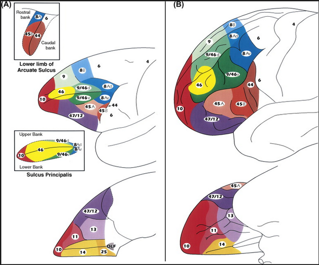 Fig 1 - Architectonic of human and monkey brain - M. Petrides (3) As you can see, the prefrontal cortex of the monkey is more elongated than the human one. It is divided in five parts: orbitofrontal (area 11, 13, 14), ventrolateral (47/12, 45, 44), dorsolateral (46, 9/46), posterior prefrontal (8), and frontopolar (10) areas (fig 1). The main role of the prefrontal cortex is to plan and coordinate our activities over time. The prefrontal cortex is an important part of brain, which is more developed for the human being and the monkey, in comparison with other animals. Nevertheless, the monkey prefrontal cortex display an homologous organization than the human brain, i.e. all cytoarchitectonic areas are present in both species (Petrides 1994)(4). As you understand, I do my internship in Neurobiology in Laboratory Stem-cell and Brain Research Institute !
During my internship, I had to focus on the the dorsolateral (46 and 9/46 areas) zone of prefrontal cortex in monkey, more particularly on the 46 area. This area extends in the dorsal (46d) and the ventral (46v) bank of the sulcus principalis (fig.1-A). The aim of my work is to identify the connectivity profiles of these two areas. The fMRI resting state - a method of neuroimaging. This method has be chosen by my host laboratory to assess the functional connectivity of 46d and 46v in monkeys with an light anesthesia. So during my internship, I should do analysis of these resting-state fMRI data with a software named AFNI . In order to identify the connectivity pattern of 46v versus 46d area. And possibly found answers (the monkey of Tamara ) ….. or maybe not :) ! |
Search the site...
Photo used under Creative Commons from DJANDYW.COM & DJANDYW.TV AKA ANDREW WILLARD
