|
The difference between the electrocardiograph and the Holter
Some cardiac disorders diagnosis with the Holter Starting from several basic characteristics of a normal ECG [7] [8], it is possible for doctors and cardiologists to detect multiple pathologies that show specific parameters and electrocardiogram models. Atrial fibrillation (AF) This pathology corresponds to a heart activity problem characterized by a fast and non-regular auricles beat leading to an irregular contraction of ventricles. It is a common disorder especially for elderly people and up-to-60 years people more likely to appear in people with other cardiac problems (hypertension, coronary artery disease …), with previous heart surgery but also lung disorders, obesity and high alcohol consumption. [9] [10] [11] 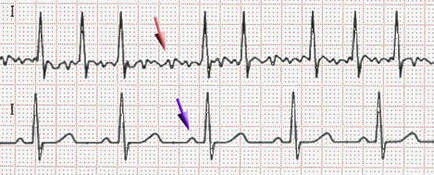 ECG in AF (at the top) and normal conditions (at the bottom). Purple arrow indicates the position of the P-wave and red arrow some shivers. We can notice that the heart beating is irregular and non-coordinated, P-wave (auricles contraction) is missing in the recording. From Wikipedia. Ventricular fibrillation (VF) This pathology refers to a heart rhythm irregularity localized on the ventricles. Instead of realizing a normal beating behaviour, the heart undertakes shivers due to myocardium fibers contractions disorder since they each have their own electrical activity (they are not contracting at the same time). Ventricular fibrillation manifests through a sudden cardiac arrest and a loss of consciousness. In the absence of treatment (cardiopulmonary resuscitation and defibrillation), complications happen leading to death making ventricular fibrillation as a serious cause of cardiac arrest (around 10%). It is more likely to happen with diseased hearts and cardiac diseases predisposition (cardiomyopathy, blood flow, coronary heart disease, valvular heart disease …), drugs overdose or sepsis. [12] [13] [14] [15] 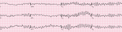 ECG in ventricular fibrillation. It shows fast and irregular oscillations of varying amplitude. No P waves, QRS complexes, or T waves are identifiables. From Wikipedia. Ventricular tachycardia (VT) Also know as V-tach, it consists in an abnormal fast heart rate localized on the ventricles. Normally initiated at the sinus node of the heart, electrical signals start in the ventricles from new autonomous cells gathered on what we call the ectopic pacemaker [16]. This region then causes premature ventricular contractions (or PVCs) [17] which can be very worrying in the case of at least three consecutive ones. Moreover, tachycardia implies a substantial heart activity with at least 100/120 beats per minute (bpm) and even 250 bpm in the framework of cardiac complications like sudden cardiac death. Due to this very fast heart activity, blood flows and pumping are critically reduced leading to very low brain and the body oxygenation. Some symptoms are known like lightheadedness, palpitations, fainting, chest pain just like the main causes which are mainly about cardiac disease predisposition (cardiomyopathy, blood flow, coronary heart disease, previous heart attack), electrolyte imbalance, anxiety, overdose or drugs. [18] [19] [20] 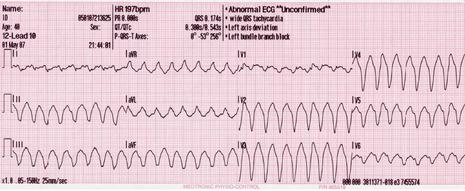 ECG in ventricular tachycardia. It shows fast oscillations with large QRS complexes and dissociated P waves which have their own rhythm. From Wikimedia Commons. 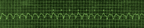 ECG in ventricular tachycardia with dissociated auricles. It shows large QRS complex and slower, regular and independent P waves from auricles (marked with dashes). From sante.ujf-grenoble.fr. Torsades de pointes “Twisting of the points” in english, this phenomena is an abnormal heart rhythm under the form of polymorphic tachycardia easily visible on an ECG. It generally appears following long-QT syndrome and starts with a phase of premature ventricular extrasystoles. If non-treated, the pathology can degenerate into ventricular fibrillation (VF) and at long-term in cardiac death. 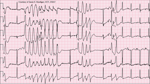 From Wikipedia. REFERENCES [1] ‘Special ECG, Holter, Formation à « l’ECG de A à Z » Par Pierre Taboulet’. Accessed 22 August 2017. http://www.e-cardiogram.com/ecg-lexique_theme.php?id_th=2&id_lex=146&PHPSESSID=47c4a327a3fd37f65869199137e327e2. [2] ‘ECG filter, Formation à « l’ECG de A à Z » Par Pierre Taboulet’. Accessed 22 August 2017. http://www.e-cardiogram.com/ecg-lexique_theme.php?id_th=2&id_lex=191&PHPSESSID=3028abdc64531925627b0c2817367a86. [3] ‘Electrocardiograph, Formation à « l’ECG de A à Z » Par Pierre Taboulet’. Accessed 22 August 2017. http://www.e-cardiogram.com/ecg-lexique_theme.php?id_th=2&id_lex=155&PHPSESSID=47c4a327a3fd37f65869199137e327e2. [4] ‘Électrocardiographie’. Wikipédia, 6 August 2017. https://fr.wikipedia.org/w/index.php?title=%C3%89lectrocardiographie&oldid=139534512. [5] García‐Niebla, J., Llontop‐García, P., Valle‐Racero, J. I., Serra‐Autonell, G., Batchvarov, V. N., & De Luna, A. B. (2009). Technical mistakes during the acquisition of the electrocardiogram. Annals of Noninvasive Electrocardiology, 14(4), 389-403. [6] ‘Holter cardiaque’. Wikipédia, 8 June 2017. https://fr.wikipedia.org/w/index.php?title=Holter_cardiaque&oldid=138018882. [7]‘Electrocardiography’. Wikipedia, 22 August 2017. https://en.wikipedia.org/w/index.php?title=Electrocardiography&oldid=796640800. [8] ‘Sémiologie et Pathologie Cardiovasculaires - L’Eléctrocardiogramme - Service de Cardiologie Du CHRU de Grenoble’. Accessed 17 August 2017. http://www-sante.ujf-grenoble.fr/SANTE/CardioCD/cardio/chapitre/301.htm. [9] Magnani, J. W., Hylek, E. M., & Apovian, C. M. (2013). Obesity Begets Atrial Fibrillation. Circulation, 128(4), 401-405. [10] Tonelo, D., Providência, R., & Gonçalves, L. (2013). Holiday heart syndrome revisited after 34 years. Arquivos brasileiros de cardiologia, 101(2), 183-189. [11] Abed, H. S., & Wittert, G. A. (2013). Obesity and atrial fibrillation. Obesity Reviews, 14(11), 929-938. [12] ‘Sepsis’. Wikipedia, 18 August 2017. https://en.wikipedia.org/w/index.php?title=Sepsis&oldid=796087587. [13] Cobb, L. A., Fahrenbruch, C. E., Olsufka, M., & Copass, M. K. (2002). Changing incidence of out-of-hospital ventricular fibrillation, 1980-2000. Jama, 288(23), 3008-3013. [14] Mogayzel, C., Quan, L., Graves, J. R., Tiedeman, D., Fahrenbruch, C., & Herndon, P. (1995). Out-of-hospital ventricular fibrillation in children and adolescents: causes and outcomes. Annals of emergency medicine, 25(4), 484-491. [15] Isner, J. M., Estes III, N. M., Thompson, P. D., Costanzo-Nordin, M. R., Subramanian, R., Miller, G., ... & Sturner, W. Q. (1986). Acute cardiac events temporally related to cocaine abuse. New England Journal of Medicine, 315(23), 1438-1443. [16] ‘Ectopic Pacemaker’. Wikipedia, 12 March 2017. https://en.wikipedia.org/w/index.php?title=Ectopic_pacemaker&oldid=770003753. [17] Osmosis. Premature Ventricular Contraction - Causes, Symptoms, Diagnosis, Treatment, Pathology, 2017. https://www.youtube.com/watch?v=wCC2bmBkpSo. [18] Bigger, J. T., Fleiss, J. L., Rolnitzky, L. M., & Groupab, T. M. P. I. R. (1986). Prevalence, characteristics and significance of ventricular tachycardia detected by 24-hour continuous electrocardiographic recordings in the late hospital phase of acute myocardial infarction. The American journal of cardiology, 58(13), 1151-1160. [19] Watkins, L. L., Blumenthal, J. A., Davidson, J. R., Babyak, M. A., McCants Jr, C. B., & Sketch Jr, M. H. (2006). Phobic anxiety, depression, and risk of ventricular arrhythmias in patients with coronary heart disease. Psychosomatic Medicine, 68(5), 651-656. [20] Chudin, E., Goldhaber, J., Garfinkel, A., Weiss, J., & Kogan, B. (1999). Intracellular Ca 2+ dynamics and the stability of ventricular tachycardia. Biophysical journal, 77(6), 2930-2941. Other sources
0 Comments
Leave a Reply. |
Search the site...
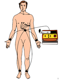
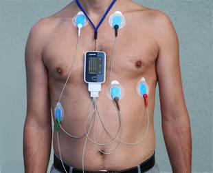
Photo used under Creative Commons from DJANDYW.COM & DJANDYW.TV AKA ANDREW WILLARD
