|
There is a lot of answer to this question. However, I can give you one. Once upon a time, in 1905, Campbell described the cytoarchitecture of the human cerebral cortex (1). However, this was a gross description in which he divided only a few general regions. At the same time (1905), Brodmann described the first map of the monkey (cercopithecus) cerebral cortex (2). To ensure a continuum between monkey and human cytoarchitectonic organization of the cerebral cortex, Bromann then described in 1909 the cytoarchitectonic organization of the human cerebral cortex using the same criteria in both species. This map is now the most used in neuroimaging studies, which refer to “Brodmann’s areas”. However, discrepancies remained between the cytoarchitectonic organization of the cerebral cortex in human and monkey. To clarify this question, Dr Petrides, a Canadian researcher, did a detailed analysis of the cytoarchitectonic organization of the frontal cortex in both species (fig 1). 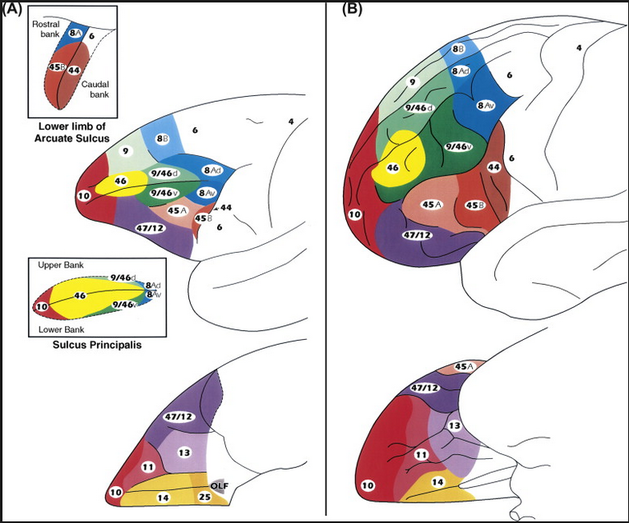 Fig 1 - Architectonic of human and monkey brain - M. Petrides (3) As you can see, the prefrontal cortex of the monkey is more elongated than the human one. It is divided in five parts: orbitofrontal (area 11, 13, 14), ventrolateral (47/12, 45, 44), dorsolateral (46, 9/46), posterior prefrontal (8), and frontopolar (10) areas (fig 1). The main role of the prefrontal cortex is to plan and coordinate our activities over time. The prefrontal cortex is an important part of brain, which is more developed for the human being and the monkey, in comparison with other animals. Nevertheless, the monkey prefrontal cortex display an homologous organization than the human brain, i.e. all cytoarchitectonic areas are present in both species (Petrides 1994)(4). As you understand, I do my internship in Neurobiology in Laboratory Stem-cell and Brain Research Institute !
During my internship, I had to focus on the the dorsolateral (46 and 9/46 areas) zone of prefrontal cortex in monkey, more particularly on the 46 area. This area extends in the dorsal (46d) and the ventral (46v) bank of the sulcus principalis (fig.1-A). The aim of my work is to identify the connectivity profiles of these two areas. The fMRI resting state - a method of neuroimaging. This method has be chosen by my host laboratory to assess the functional connectivity of 46d and 46v in monkeys with an light anesthesia. So during my internship, I should do analysis of these resting-state fMRI data with a software named AFNI . In order to identify the connectivity pattern of 46v versus 46d area. And possibly found answers (the monkey of Tamara ) ….. or maybe not :) !
10 Comments
Yulian
23/3/2017 08:11:21 am
Hello Isa, thanks for sharing this topic, I want to know more about it! Tell me... how can you see connectivity between the different brain regions with the fMRI? :)
Isabelle
5/4/2017 05:28:33 am
Tamara's monkey is the monkey on the trip to Bidar! The monkey represents an answer found by surprise.
Yulian
23/3/2017 08:11:55 am
And could you also explain what is the issue with the Monkey of Tamara?
margaux
31/3/2017 01:35:35 am
Tamara has a monkey?
Yu
1/4/2017 08:35:09 am
I don't know .. but Isa talk about a Tamara monkey on her blogpost...
Margaux
31/3/2017 01:46:17 am
Hi Isa!
Isabelle
13/4/2017 03:12:06 am
Hello Margaux ! Thank you for your questions ! :)
Régine
31/3/2017 04:16:57 am
Hi Isa!
Isabelle
5/4/2017 05:43:05 am
According to the theory, the dorsal bank is specific to the functional activity of the movement and the ventral bank is related to the recurrence of the movement. The area 46 is the center according to the cognitive researchers and is separated into two very distinctive parts by the sulcus principalis (dorsal and ventral). So our objective is to find the difference between these two banks in region 46. To find perhaps a distinction in order to verify this theory .
Cécile
1/4/2017 08:38:00 am
Hi Isa! Leave a Reply. |
Search the site...
Photo used under Creative Commons from DJANDYW.COM & DJANDYW.TV AKA ANDREW WILLARD
