|
During my internship, I write and adapt cards about some diseases for patients. These card inform a patient about his or her disease difficulties, especially when the disease is severe and has huge consequences on the patient’s quality of life. This it is necessary to help him or her to be aware and active during the treatment. At the beginning of my internship, I have been asking myself: : what should be the content of the cards? how much can I write on it ? ? How can I write it ? From these questions, I started my research with a law perspective. I found an interesting answer to a part of my questions about what should be the content of the card. It was interesting to study the evolution of the informed consent because this evolution illustrates the need of the patient through his or her complaints during the time. “The old Hippocratic ethic saw the patient as a weak, debilitated, childlike victim, incapable of functioning as a real moral agent…” - Robert Veatch Three crucial evolutions have been made by the Court of Cassation in France about the implication of doctors in the process of information :
Consent of the patient was a subject of interest since 1889 [4], when the surgeon could be incriminated because the patient was not informed about the different existing therapies. In May 20th 1936, the Court of Cassation established as a contract the nature of the relation patient-physician [4]. This contract engages the practitioner to get the consent of the patient. A consent required enough information for the patient to decide in all good conscience [4]. Thus, at this point the doctor was already engaged to let the patient have all the information to take his decision. However, the amount of information considered sufficient to take a decision was not yet established and are different depending of the place and the period. October 7th 1940 was the beginning of the Conseil de l’Ordre des Médecins (Council of the Order of Physicians) during the Vichy government. If the creation of this council was thinking before this date, Vichy government used this opportunity to create discriminatory rules against Jewish patients. This institution was completely changed by the new government in 1945. The professor Portes decided to write the Code de Déontologie Médicale (Code of Medical Ethics) in 1947 to avoid this situation and have a practice of medicine ethic and moral. This Order was “responsible for the maintenance of morality, probity and devotion crucial for the practice of medicine” [5]. Professor Louis Portes, in 1950, declared that “emotion or pain dominate his [patient] drive so his will is based on nothing concrete” [6]. Thus, the model patient-physician continue to be “paternalistic” during this period. Actually, this code continues to be used but it is updated according to the evolution of French society. Simultaneously, a driver was victim of an accident in 1930 and his surgeon decided to practice an osteosynthesis without the informed consent. This operation had catastrophic side-effect for the patient that lost his arm. M. Teyssier, the driver, engaged liability lawsuit against his surgeon because the surgeon had decided the operation for his own interest and without informing the patient about the risks and the possible alternatives of treatments. Teyssier judgement, pronounced January 28th 1942, forbids the surgeon and confirms the duty for a doctor to let the patient be informed before taking a decision [7]. In 1951, M. Birot was amputated after an error of diagnostic. He engaged liability lawsuit and one of the arguments was the lack of information given by the practitioner that caused a non-informed consent for the surgical intervention [8]. However, the May 29th 1951, the Court of Cassation decided that : “when [the patient] consents to the operation in a moment of clarity, he has to report the proof that the surgeon neglects his duty because of a lack of information about the real implication of the operation” [8]. This decision was contested because it engages the patient to the proof that the doctor misses an information while he doesn’t have the medical knowledge and he is in a position of vulnerability. Shifting the burden of proof of information occurs in 1997, when the Court of Cassation decides to cancel the judgement from Rennes’ Court of Appeal. This judgement rejects the application of a patient victim of a colonoscopy complications. The patient, M Hédreux, defends the non-information of the risk of this examination. The first judgement decided that the patient has no way to proof the lack of information. However, the Court of Cassation, The February 25th 1997, decided that: ” the physician is attempt to inform his patient, so he must proof that he fulfils his duty” [9]. Thus, the patient is free from the obligation to demonstrate that he was well informed. This obligation is now for the doctor. However, it is important to notice that nothing indicates what type of proof the doctor have to give. Or even, the quality or the quantity of information was not detailed yet. If a doctor does not have to convey his patient to practice something that the patient doesn’t want [10], he must inform his patient about all the risks that an operation could cause (except if the patient doesn’t want to know or in a case of an emergency) [11]. In others words, they mean all the serious risks that could create an investigation or a treatment. This responsibility is not “excuse just because this risk is exceptional “ (Cassation court, judgement established the 07/10/98) [12]. All these laws were established in order to get an informed consent from the patient to an operation, an examination or a treatment. They are aligned with the Code de Déontologie Médical, in particular with the articles 35 and 36. The article 35 states that: “The physician owes a loyal, clear and appropriate information on his health, the investigations and the care which he proposed to the patient” [13]. These articles and different laws are supported by the chapter II article 11 of the law n°2002-303 establish the March 4th 2002 about sick peoples rights and system health quality [14]. This article is called “healthcare system users information and expression of their will” [15]. Before a hospitalization, a patient can ask La charte des personnes hospitalisées (charter of hospitalized person) which describes all his rights. The two major principles about information are : - accessible and loyal information for the patient (for the investigation, treatment, prevention act, potential alternative) [16]; - a medical action can’t be done without the free and informed consent [17]; To conclude, this study depicts the evolution of law on patient’s information. During the history, two majors changes have been done: informing the patient is necessary and doctor have to proof that they have informed their patients before any medical act. These changes deeply transform the relationship between the patient and the physician. Now, the patient is not approached as a vulnerable child who can’t be the actor of his own healthcare but he is considered as a person capable to evaluate and understand the information in order to take a decision. However, this relationship is suffering because of the proof and the relativity of some laws. No answer was supplied by law about how communicate the information and when. Some practitioners are afraid to be legally pursued because of a lack of information. This fear can interfere during the process of information and harm the quality of the interaction which could be difficult for both patients and practitioners because of the significance and importance of the information to deliver. Bibliography : [1] : Déontologie médicale de Max. Simon (1845), Bernard Hoerni La Revue du Praticien,vol. 64 1474-1477, Décembre 2014 [2] : Déontologie Médicale ou Des devoirs et des droits des médecins dans l’état actuel de la civilisation, Docteur Max Simon, 1845, J.B. Baillière, translated from french : “doit peser chacune de ses paroles, dans la crainte qu’un mot imprudent ne révèle au malade l’affection grave dont il est atteint” [3] : Déontologie Médicale ou Des devoirs et des droits des médecins dans l’état actuel de la civilisation, Docteur Max Simon, 1845, J.B. Baillière, translated from french : “il doit respecter avec la même réserve la dernière espérance qui attache l’homme à la vie. “ [4] : http://www.univ-reims.fr/gallery_files/site/1/90/1129/1384/1536/1577/1579.pdf [5] : M Billoux, Health Minister in 1945, translated from French : « chargé du maintien des principes de moralité, de probité, et de dévouement indispensable à l’exercice de la médecine… et à l’observation des règles dictées par le code de déontologie » [6] : Introduction générale à la bioéthique : histoire, concepts et outils, Guy Durant, coll. “Fides”, 2005, translated from French : “[...] que son affectivité est dominée par l’émotivité ou par la douleur et que sa volonté ne repose sur rien de solide” [7] : Arrêt Teyssier de la Cour de Cassation, 28 janvier 1942 : quelques remarques sur une décision “oubliée”, Bernard Hoerni et J. P. Bouscharain http://www.biusante.parisdescartes.fr/sfhm/hsm/HSMx2001x035x003/HSMx2001x035x003x0299.pdf [8] : translated from french : “il appartient toutefois à celui-ci lorsqu’il se soumet en pleine lucidité à l’intervention du chirurgien, de rapporter la preuve que ce dernier a manqué à cette obligation contractuelle en ne l’informant pas de la véritable nature de l’opération qui se préparait” https://www.pedagogie.ac-aix-marseille.fr/upload/docs/application/forcedownload/2012-06/dgda98en.pdf [9] :translated from french : "médecin est tenu d’une obligation particulière d’information vis à vis de son patient et qu’il lui incombe de prouver qu’il a exécuté cette obligation" https://www.lamedicale.fr/documents/201008infomdc.pdf [10] : https://www.legifrance.gouv.fr/affichJuriJudi.do?idTexte=JURITEXT000007409555 , Court of Cassation 18/01/2000 [11] : https://www.legifrance.gouv.fr/affichJuriJudi.do?idTexte=JURITEXT000007038714, Court of Cassation 07/10/1998 [12] : Cass 07/10/1998 translated from french : “il n’est pas dispensé de cette obligation par le seul fait que ces risques ne se réalisent qu’exceptionnellement” [13] : code de déontologie médical 2017 [14] : https://www.legifrance.gouv.fr/affichTexte.do?cidTexte=JORFTEXT000000227015&categorieLien=id, Loi n° 2002-303 du 4 mars 2002 relative aux droits des malades et à la qualité du système de santé (1) [15] : translated from Loi n° 2002-303 du 4 mars 2002 : “information des usagers du système de santé et expression de leur volonté” [16] : translated from Charte des personnes hospitalisées :“ L’information donnée au patient doit être accessible et loyale” [17] : translated from Charte des personnes hospitalisées : “un acte médical ne peut être pratiqué qu’avec le consentement libre et éclairé du patient”
3 Comments
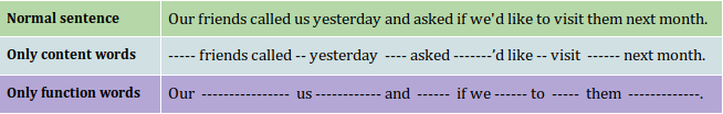 Table 1. Sentence with function words and content words deleted. We can observe that, while when function words are deleted we can still understand the meaning of the sentence; when content words are deleted we can not. Function words can also help us categorise content words in their different categories (noun, verb, adjective and adverb). For example, if I say “she blickets”, you will classify ‘blickets’ as a verb, however if I say “the blickets” you will classify ‘blickets’ as an object now. This shows us that the choice of function words creates a syntactic context that helps us categorise novel content words (Bernal et al., 2007; Shi and Melançon 2010; de Carvalho et al., 2016). This process is called syntactic bootstrapping (Gleitman, 1990).
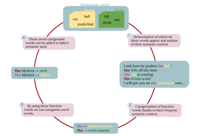 Figure 1. Semantic Seed Hypothesis. By using the semantic seed and some memorised context where this semantic seed appears, the baby can get to know some function words. Then, they will be able to relate these function words to syntactic contexts that will help them to categorise novel words. More precisely, the experiments we are running at the BabyLab (LSCP), in which I take part during my internship, try to test the validity of the Semantic Seed Hypothesis. To know how do we do it, read my next blogpost!
References: Bernal, S., Lidz, J., Millotte, S., & Christophe, A. (2007). Syntax Constrains the Acquisition of Verb Meaning. Language Learning and Development, 3 (4), 325–341. http://doi.org/10.1080/15475440701542609 de Carvalho, A., Lidz, J., Tieu, L., Bleam, T., & Christophe, A. (2016). English-speaking preschoolers can use phrasal prosody for syntactic parsing. Journal of the Acoustical Society of America, 139(6), EL216-EL222 Christophe, A., Dautriche, I., de Carvalho, A., & Brusini, P. (2016). Bootstrapping the Syntactic Bootstrapper. In Proceedings of the 40th annual Boston University Conference on Language Development. Boston, MA: Cascadilla Press, 75-88. Gleitman, L. (1990). The Structural Sources of Verb Meanings. Language Acquisition, 1, 3–55. Shi, R., & Melançon, A. (2010). Syntactic Categorization in French-Learning Infants. Infancy, 15(5), 517–533. http://doi.org/10.1111/j.1532-7078.2009.00022.x At the Astrobiology department within the LISA, an Atmospheric Systems Laboratory. (LISA – Laboratoire Inter-universitaire des Systèmes Atmosphériques) Créteil, France. The end of my internship is fast approaching and I have some ideas of future improvement for this program. However, I would maybe not have time to set up these new ideas. Let me introduce some articles which can bring new informations.
1. Polycyclic aromatic hydrocarbon - Polycyclic aromatic hydrocarbon processing by cosmic rays (Micelotta, Jones and Tielens, 2010) Actually, any radiolysis destruction constant under GCRs radiation was determined. This article presents the lifetime of PAHs under GCRs radiation. A theoritical model determine that the lifetime of PAHs under GCR radiation depends of the molecule size, on the electronic excitation energy E0 and on the amount of energy available for dissociation. According to their estimation, the lifetime of PAHs under GCR radiation is between 107 years for the smallest molecules (~10 C-atoms) and 109 years for the larger molecules (~1000 C-atoms). However, our program simulate the survival of molecules during few years and could not take into account a destruction during 107 years. We are, thus, waiting for an experimental radiolysis constant which might be determined thanks to the return of the spatial mission “EXPOSE” which exposes organic matter to a galactic environment on the International Space Station or wainting for a laboratory experiment which can give us this radiolysis constant. 2. Mars sedimentary rock erosion rates - Mars sedimentary rock erosion rates constrained using crater counts, with applications to organic-matter preservation and to the global dust cycle (Kite and Mayer, 2017) This article presents the Mars sedimentary rock erosion rates and the consequences on the preservation of organic matter. Their conclusion were that the radiolysis is less of a threat to the preservation of ancient, complex organic matter. Therefore, the exhumation rate at the paleolake deposits in SW Melas Chasma is relatively high. Whereas, radiolysis is more of a threat to the preservation of ancient, complex organic matter when the exhumation rate is relatively low such as at the Oxia Planum and Aram Dorsum. However, the mean value of their exhumation rate was equal to 100 nm/yr, which is too low to have an impact on our model. 3. “Solar Energetic Particles” (SEPs) or called also “Solar Cosmic Rays” (SCRs) - Degradation of the organic molecules in the shallow subsurface of Mars due to irradiation by cosmic rays (Pavlov et al., 2012) - Modelling the surface and subsurface Martian radiation environment: Implications for astrobiology (Dartnell et al., 2007) These articles discuss about impact of “Solar Energetic Particles” (SEPs) or called also “Solar Cosmic Rays” (SCRs) . (Pavlov et al., 2012) estimated the SEPs flux at the Mars orbit was assumed to be 33 protons/cm2/sec. SCRs particles were assumed to have energies in the range of 1 MeV – 1GeV. SCRs particles with energies below 1 MeV would be absorbed in the atmosphere and would not reach the Martian surface. There are very few SCRs particles with energies above 1 GeV. However, the SEPs flux is dependent on the 11-year solar activity cycle. Then, the impact of SEPs on organics could be almost easly model at the surface of Mars. 4. Adenine on the Martian Surface under secondary particles - Degradation of Adenine on the Martian Surface in the presence of Perchlorates and Ionizing Radiation: A Reflection Time-of-flight Mass Spectrometric Study (Gobi, Bergantini and kaiser, 2017) This article describe the radiolysis decomposition of adenine (C5H5N5) under Martian conditions irradiated with energetic electrons that simulate the secondary particles originating from the interaction of the galactic cosmic rays (GCRs) with the Martian surface. Neat adenine and adenine-magnesium perchlorate mixtures were experimentaly irradiated. Then, the destruction of Adenine under secondary particles on Mars could be added to the model with a new type of radiation. Moreover, thanks to the set-up MOMIE (Mars Organic Matter Irradiation and Evolution) developed at the LISA laboratory, the photodestruction constant of Adenine caused by UV radiation was already determined and during my previous internship in this lab, I was involved in the experiment to study the chemical evolution of Adenine under UV radiation in martian conditions (temperature and pressure). Throughout L2, our class worked on the replicability of experiences with the AJA project. Redoing someone’s protocol and analyzing the obtained results helped us see the importance of being able to replicate an experience both to verify a method and to verify conclusions. To be able to accurately replicate an experience, a detailed protocol is needed. To maintain traceability of experiments in a lab, you are required to keep an updated lab notebook that stays within the team. Why is it important to keep a good lab notebook? First of all, keeping an updated and clean lab notebook is important for you. It helps you move forward productively in your experiments, to make sure you don’t redo the same experiment or reproduce the same mistakes. It can also help you explain your results or justify changes in methodology for example. Secondly, your lab notebook will be used by others after you leave the lab. This detailed trace of your work helps others save time both in understanding what has been done before and why it was done. What is a good lab notebook (according to me)? Objective: why you are doing this experiment, Protocol: detailed protocol with specific material used, with added comments of things that changed, any things you noticed during the experiment, Results: detailed results with labelled figures, highlight anything you find interesting Conclusions: what you can conclude from this experiment, and the next steps based on this Why do I care?
I based the beginning of my work a lot on the work of previous interns, so when details were missing, it was complicated to proceed. I needed to know what primers had been used, and a specific trace of exactly what sequence was used was not always present. Also, choices linked to primer pairs were not always justified. One experiment was done with several different options and directly after only one pair was used, so I had to redo this experiment in order to understand if there was a problem in efficiency. What can seem really clear in your head might not be for someone else, so writing down all the details in your lab notebook is really important! I received an interesting question on my last blog post so I'd like to answer it. I was explaining that I had to increase the concentration of the primary antibody when doing my immunostaining to make sure that the protein I'm studying will be marked. I have been asked whether this could increase the background noise (which was already pretty high!). Well, I think this is a good point and usually the optimal dilution for the antibody is determined by running a serial dilution test. This should allow to find the best signal to noise ratio. If you look at the picture I got this morning, the background noise is effectively very high and there is no signal specific for the protein, which means that increasing the concentration of the primary antibody was not optimal. 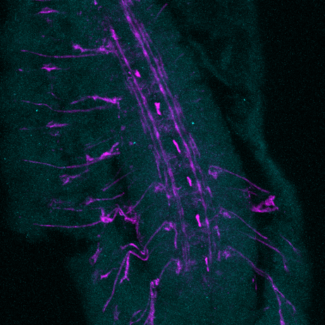 Fungal organisms are commonly exploited by the human activity: the production of antibiotics, vitamins, enzymes and other metabolites deserve pharmaceutical, food and biotechnological industries. Fungi have a major place on the ecological dynamics of ecosystems by using different roles, such as symbionts (such as lichens, mycchorhize…), parasites, pathogenic agents and saprophytes, with a degradation activity that permit the transfer of nutrients from the degraded organisms to soils and water. In this study, we will focus on the fungal decomposer community. Here, we will focus on filamentous species, that, contrary to yeasts, are multicellular; however, this distinction is relative, considering dimorphic fungi that may take both forms. The fungus-like organism distinction concern mainly Oomycota, a rein of organisms which has similar feature with fungi but present differences, such as a cellulose cell wall instead of chitin, that made them similar to plants. Fungi, and fungus-like organisms are eukaryotic organisms. Filaments of fungi are also name hyphae (singular: hypha)(1). The filamentous fungi growth is apical: the extreme part of filaments, is the place where cellular division occurs. The hyphae network is called mycelium. A cytoplasmic flow occurs to the hyphal tip, wearing secreted vesicle to the apex. At this position, the wall is thinner . Some fungi are septate, meaning that septa, or cross-walls, are present at regular intervals. Woronin bodies appears close to the cross-walls pores. These are membranous organelles that might close pores when a compartment is damaged, became too old or undergo differentiation. Septa present pores, where cytoplasm and even the nuclei can pass. Hyphae are not made with cells but by interconnected compartments. At the oldest part of the mycelium, present vacuole that may take most of the space on the hypha, restricting the cytoplasm, the nucleus and other organism to a small fraction. When hyphae are too old, compartments may be empty, closed from the rest of the organism. Cells walls are digested by enzymes and the rest is used by the mycelium to produce a thick-wall resting spore, named chlamydospores. The mycelium growth is multidirectional, with several order of branches, and interconnections between branches. Fungi has no predetermined age, size, or shape : they are spatially and temporary undetermined. The cell-wall is composed of Beta-glucan polysaccharide for rigidity, chitin or cellulose (oomycota), with mannoprotein that permit external exchange of molecule; and ergosterol, a steroid that take place of cholesterol of animals, and make the layer more fluid.  Septate of a hypha with its semi-compartments. Source : http://bugs.bio.usyd.edu.au/learning/resources/CAL/Microconcepts/Diversity/fungi.html The nutrition of fungi is chemoheterotrophic. Its solubilize nutrients with external enzymatic catalysis and then absorb nutrient that cannot enter into the organism without depolymerization. . The growing mycelium is normally always attached to the food source, and the shape of the culture is influenced by the need to obtain nutrients. Enzymes are released in the zone of erosion of the hypha that permit the secretion of large molecules (from 20 to 60.10^3 Da). On this located area, fungi is weaker and may separate from the mycelium if its threaten by an external factor. Polymer-degrading fungi may also locally synthesize antibiotics in the closest area of nutrient uptake, probably to compete with other organisms. This has be reported in the Rhizoctonia spp. By Burton and Coley-Smith (1993). Usually, the carbon source differ with fungal strains, from simple methane to larger complex macromolecules, such as lignin, keratin, cellulose... For common laboratory fungal culture, the use of potato-dextrose, malt or cornmeal agar is widespread. These culture media are more acidic than those for bacteria; and contain relatively more nitrogen. Complementary nutrients may by added for appropriate culture such as vitamins, asparagine (as a source of nitrogen for strains that cannot use nitrate, or ammonium), or other more specific nutrients. Spores production is part of the reproduction of fungi organisms: this aspect matter concerning laboratory security rules. Spores are formed in the sporangium, a large multinuclear cell that secrete spores by cytoplasm separation. Some fungi lack a sporangium, and produce spores by several phenomenon, budding, fragmentation… Major taxonomic groups of fungi and fungus like organisms include Chrytridiomycota, Zygomycota, Ascomycota, Deuteromycota and Oomycota. They differs in the sexual reproduction. For example, the life cycle and sexual reproduction of Mucor, a specie employed in bioremediation process that belong to the Zygomycota group, start with an asexual reproduction, with a production of sporangiophores . Spores are released, then fix and germinate to a somatic hyphae. The mating occurs when complementary types of aerial branches of hyphae growth towards each other, mate, produce a progametangia that separate from each other. The final gametangia will fuse, and create a diploid compartmented zygospore. Then, the meiosis occurs and produce a new hypha. 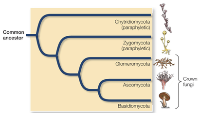 Source : Life, the Science of Biology, Eight Edition, 2007. The fungal kinetic growth present thew wide-known model: after the inoculation in laboratory, first occurs a lag phase, followed by an exponential growth, that then decelerate until becoming stationnary, and finally and autolysis phase. On laboratory condition, when a culture is saturated, the organism start to sporulate; spore may spread and contaminate other non-sterile experiment, but can also threaten the human health while breathing. Source : Introduction to Modern Mycology, Blackwell Sciences - J.W. Deacon (1997)
As you may know, I am trying to understand how Lola, a transcription factor expressed in early born neurons, can repress the expression of stem cell genes to prevent the neuron to neuroblast reversion. Lola interact with the Rcd5 protein which recruits the transcription machinery Previously, the lab I working on did a yeast two hybrid assay (you can read my blog post about it) and discovered that Lola might interact with Rcd5. Interestingly, Rcd5 has been shown to facilitate the recruitment of RNA Polymerase II preinitiation complexes (i.e. the transcription machinery) to the promoter regions of target genes (Anderson et al, 2010). The protein promotes genes transcription required for normal proliferation. Localizing the Rcd5 protein using immunostaining and confocal microscopy In order to localize the Rcd5 protein in the Optic Lobe, where Lola is required to maintain immature neurons differentiated, I stained wild type Drosophila larve brains. I used mouse the anti-Dlg antibody because Dlg is a marker of membranes and should indicate the shape of the brain. It is stained in blue in the figure. Rat anti-Elav is stained in red. Elav is a transcription factor specifically expressed in early-born neurons. Finally, guinea pig anti-Rcd5 (obtained from Anderson’s lab) stained in green shows the protein I am interested in localizing. I used a confocal microscope to obtain images, with a 40x and 63x magnification. As you can see, it is not clear that Rcd5 (in green) is preferentially expressed in the neuroblasts (big dark dots).
What’s the best between staining larve brain and embryo? So I repeated the staining in embryo of wild type Drosophila, because immunostaining are usually more efficient than in larve brains. I also tried different concentration for the anti-Rcd5 antibody: 1:500 because Anderson et al used this concentration and a higher concentration of 1:100. However, this did not give more conclusive results and it is not clear where the protein is localized.
Reliable staining or Residual fluorescence?
Because the staining of the protein (brightness of the color) is so weak, I was wondering whether the green dots that I was observing on the picture could only be some background noise from the fluorophore. Indeed, to perform an immunostaining, a primary antibodies is used to target the protein of interest, in my case Rcd5, and a secondary antibody is used later in the process to add fluorescence. So, the green dots on the figures might only be some residual staining from the secondary antibody instead of the actual protein. This would be possible because the primary antibody, anti-Rcd5, is quite ancient now (from 2010). To test whether the anti-Rcd5 antibody is efficiently marking the Rcd5 protein, I decided to:
Although my slides are ready, I’ll have to wait until tomorrow to use the confocal microscope and know the answer… Reference: Andersen, D.S., Raja, S.J., Colombani, J., Shaw, R.L., et al. (2010) Drosophila MCRS2 associates with RNA polymerase II complexes to regulate transcription. Molecular and cellular biology. [Online] 30 (19), 4744–4755. Available from: doi:10.1128/MCB.01586-09. Why I did this post ? For this post I was looking for a subject, linked to Glowee, but without confidential informations, and of course enough interesting. I founded the idea when Simon the designer came for it morning coffee! So, I wanted to write a post on design, talking about it link with biology. So, in this post, I will not talk about the symbiosis of two organisms but the “symbiosis” between design and biology. In fact, both are important parts at Glowee, but also in other fields or companies. So, I wanted to talk a bit about design, and why these two fields are linked.  But what is design? There is no real definition of design, because it change in function of the time, the fashion, the world or the cultures. But to describe design more particularly, the AFD (Alliance Française des Designers) give this following definition: “Design is a creative, multidisciplinary and humanistic intellectual process whose goal is to deal with and solve everyday problems, small and large, linked to economic, social and environmental issues.” And if biology needs design ? and vice versa? We don’t always pay attention to the design of objects, but for many of them, someone has thought about the shape, the size or the way to use it. It is also the case in many of instruments or machine in laboratories. Design is everywhere! In Glowee, the work of the designer is useful to create new shapes of bioluminescent stickers, new ideas of decors or new kind of utilisations of bioluminescent lights. Glowee absolutely need innovation in therm of light production but also design innovations, in order to create new kinds of products for example. Still working in the lab for biological researches, I see designers talking with the lab team, about our new researches, results and also needs for the experiments. So, the biological part is also a source of ideas for the designers.
A short explanation of the biodegradation of pollutants by fungi Let’s talk about mycoremediation. Different pollutants are present in the environment, soils, water stream, air and oceans: We can consider different pollutants : polycyclic aromatic hydrocarbons, or PAH, a families of stable chemical products composed from many aromatic rings that make them really stable; pesticides, that make huge damages in the environment ; synthetic dyes, from the textiles industries, are often found in wastewaters and came from the water from your washing machine; TNT and RDX, explosives compounds used in bombs and spread spread over environment after war conflicts ; oils, fragrances and diesels form petroleum hydrocarbons; heavy metals and metalloïds; endocrine disruptors, chemicals compounds that can interact with endocrine organs and lead to a disrupt in human and animal metabolisms – theses compounds, including phtalates, phenols, organochlorides metallo-organic compounds and medicines has a wide range of origins, including pesticides, medicine and plastic micro products… In order to counteract these different compounds and their negative effect in the environment, fungi present several properties that make them suitable from remediation : They can be tolerant to extreme conditions, they developement permit them to reach chemicals pollutants, that are sometimes adsorbed to surfaces and over biomass, blocked in tiny pores, precipitated or dissolved in the water. They also don’t need these pollutants for they development. A plus for they application is that they sometimes bring waters and nutrient to the contaminated environment, permitting the revival of the ecosystem. In the family tree of fungi, most pollutant degraders belong to the phyla Ascomycota and Basidomycota, and Mucoromycotina, a subdivison of Zygomycota. 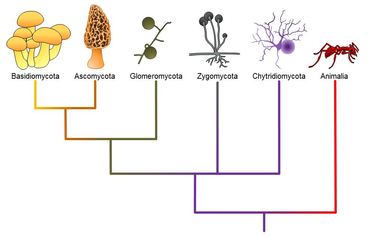 A simplified scheme of fungi phylogeny. Source : http://cnx.org/contents/uVdR6VSq@8/Kingdom-Fungi The studied fungi are filamentous fungi : unlike yeasts, they spatially growth forming a hyphae network. Because of they major role in the rhizosphere, they are able to cop with plants by absorbing some pollutants; they can also host endomycotic bacteria. Fungi generally solubilize external nutrient with enzymes before absorption. Fungal degradation is also advantageous in remediation because of the enzyme are not highly specific and can degrade different and related compounds. Its is know that some Cladophialora ans Exophiala dedrade toluene. Aspergillus and Penicillium can degrade some pesticides, TNT, PAHs and hydrocarbons. PAHs can be degraded by several (sub)phylums, such as Kickwellomycotina, Chytridiomycota, Glomeromycota… The subphylum of Mucoromycotina has been less studied concerning the degradation properties, but, the genera Cunninghamella, Mucor and Rhizopus belonging to this subphylum can degrade PAH, textile dyes and TNT. How does fungi degrade pollutants. We can divide degradation into two types : external degradation, and internal degradation. The external oxidation involve enzymes such as laccases and peroxydases, resulting in cleavages, aromatic-ring fission, oxidative-coupling products… Internal degradation happened inside the fungal organism. First, an initial attack in the cytochrome P450 enzyme permit a further treatment by pollutants, such as conjugate formation using transferase, or further catabolism. The biodegradation lead product from pollutants that act as reactants. The mineralization of pollutants can ultimaltely lead to the production of H20/CO2, due to oxidations/reductions. But generally, remediation lead to products that are less toxic, such as bound residues, metabolites excretion… Source : Untapped potential : exploiting fungi in bioremediation of hazardous chemical, Harms, Schlosser ans Wick, 2011, Applied and Industrial Microbiology.
Last week, I went for the first time on site to visit a desalination plant that the company I am working for is operating. The plant furnishes 100 MLD (Million Litres per Day) of drinkable water to the nearest city. They have been operating the plant for the past 4 years and have therefore had the time to face a lot of problems. Indeed, when the plant first started to work, it was not producing the 100 MLD but rather 70 MLD while it had been design for 100. While interacting with the director of the plant who did the trouble shooting – fact of finding where the problem is coming from in a plant –, I had the possibility to understand what were the key points to limit such difference between what it is designed for and the real outlet and therefore reinforced my knowledge to design better. I will talk about five of the points I found interesting and maybe less obvious.
The first one is the use of valves in the pipelines. Indeed, to regulate the flow valves are used but are producing a pressure loss in the incoming flow. This pressure loss will vary according to the types of valve used. When designing the plant they had used ball valves for which the pressure loss is approximately 0,5 bars but they have replaced most of this valves by butterfly valves which allow less regulation but have a pressure loss of 0,1-0,2 bars. Another point is the presence of bends in the pipes. The more bends they will be, the more pressure loss will happen and therefore the less water will be arriving to the equipment and hence produced. The director directed some work to straightened the pipe at maximum. This taught me that in order for the plant to work properly the layout should be well thought. Also, even if ideally of all the units should be closed and no water should be coming out of any of any of them it does not happen in reality and therefore drains should be designed. Sometimes, due to space requirement or a miss in the design the drains are also the pipe galleries. However, this is far to be ideal as the pipes, valves and other equipment will be damaged or corroded by the presence of water. Therefore, when designing we should separate the pipe galleries from the drains. The next problem is regarding the access to the units. When designing we might forget the on-site experience and that one should be able to get near all the units to be able to see what is happening in case of problem. Therefore, the space between units is very important. A last very important point is the selection of the pumps. We should try to limit the friction and the resistance of pumps to allow a better efficiency of such and therefore less electrical consumption and lesser operation and maintenance cost. This will also permit to reduce the flow. There are many other points which are needed to be taken into account but I will not expend myself about them as it might start be boring for you. |
|||||||||||
Search the site...
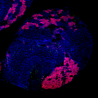
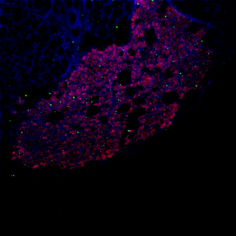
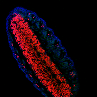
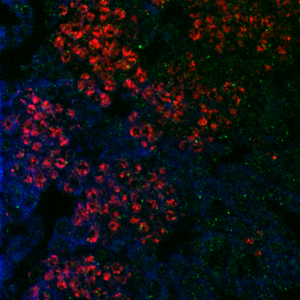
Photo used under Creative Commons from DJANDYW.COM & DJANDYW.TV AKA ANDREW WILLARD
