|
Let's begin with the basics ... ECG, or electrocardiogram, corresponds to the heart electrical activity representation made using an electrocardiograph. It is a critical tool, non-invasive, very cheap and highly usual examination used in electrocardiography to discover most of existing cardiac pathologies. [1] [2] [3] Heart scientific approach
The right part of the heart (the first pump) is dedicated to carry deoxygenated blood from the organs and muscles to lung’s capillaries to undergo gas exchanges with the external environment: O2 is recovered whereas CO2 is discharged. Afterwards, the left part of the heart (the second pump) carries the oxygenated blood directly to all body organs and muscles which will absorb it as well as nutrients to finally release wastes and CO2.
ECG functionning and principles These are these different electrical activities behaviours (in this case, their amplitude variation or electrical potential variation to be more precise) that we are trying to detect using an electrocardiograph depending on the time. Indeed, these different steps can be recognized by the different phases found in it. 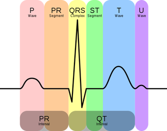 Graphical representation of a ECG from a healthy patient. P wave: auricular depolarization; QRS complex: ventricular depolarization; T wave: ventricular repolarization; PR and ST segment: latency isoelectric period. From Wikimedia Commons. Finally, we usually measure the heart activity thanks to at least two electrodes that will enable to get the heart activity under the form of an ECG. As the heart is an three-dimensional organ, the two electrodes will give us this activity under the form of a single vector called leads. Thus, depending on the position and the number of electrodes (12 maximum) placed according to the Einthoven’s triangle, we will obtain different leads and then different information about heart activity as for proper functioning as for proper morphology.
REFERENCES
[1] ‘Cours - Cardiologie - L’électrocardiogramme, Les Bases Pour Comprendre (1ère Partie)’. Infirmiers.Com, 9 April 2009. http://www.infirmiers.com/etudiants-en-ifsi/cours/cours-cardiologie-lelectrocardiogramme-les-bases-pour-comprendre-1ere-partie.html. [2] ‘Électrocardiogramme : Fonctionnement et Applications - Ooreka’. Ooreka.Fr. Accessed 16 August 2017. https://defibrillateur.ooreka.fr/comprendre/electrocardiogramme. [3] ‘Heart’. Wikipedia, 2 August 2017. https://en.wikipedia.org/w/index.php?title=Heart&oldid=793554003. [4] ‘Myocyte’. Wikipédia, 24 April 2017. https://fr.wikipedia.org/w/index.php?title=Myocyte&oldid=136753036. [5] Bonny, A., Lele, E. C. B., Mandengue, S., Larrazet, F., & Amara, W. (2013). Différences ethniques de l’électrocardiogramme entre une population de noirs africains et de blancs européens âgés de moins de 35ans. La Presse Médicale, 42(4), e96-e105. [6] Macfarlane, P. W., McLaughlin, S. C., Devine, B., & Yang, T. F. (1994). Effects of age, sex, and race on ECG interval measurements. Journal of Electrocardiology, 27, 14-19. [7] Devi, M. R., Arvind, T., & Kumar, P. S. (2013). ECG Changes in smokers and non smokers-a comparative study. Journal of clinical and diagnostic research: JCDR, 7(5), 824. [8] Murata, K., Landrigan, P. J., & Araki, S. (1992). Effects of age, heart rate, gender, tobacco and alcohol ingestion on RR interval variability in human ECG. Journal of the autonomic nervous system, 37(3), 199-206. [9] Cellina, G. U., Honour, A. J., & Littler, W. A. (1975). Direct arterial pressure, heart rate, and electrocardiogram during cigarette smoking in unrestricted patients. American heart journal, 89(1), 18-25. [10] FRIEDMAN, L. A., & Kimball, A. W. (1986). Coronary heart disease mortality and alcohol consumption in Framingham. American Journal of Epidemiology, 124(3), 481-489. [11] Evans, W. (1959). The electrocardiogram of alcoholic cardiomyopathy. British heart journal, 21(4), 445. Other sources
2 Comments
Nikola
6/9/2017 09:48:11 am
Hi :)
Corentin
10/9/2017 07:16:08 am
Hey Nikola! Leave a Reply. |
Search the site...
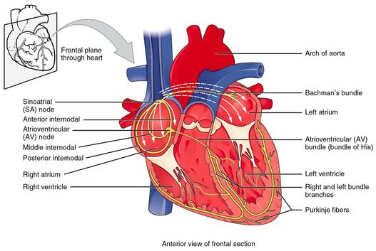
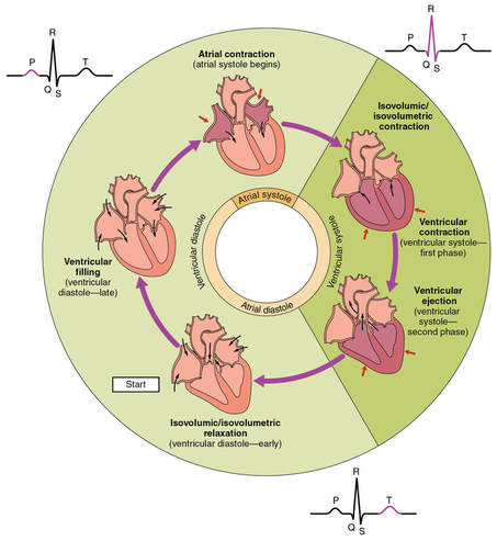
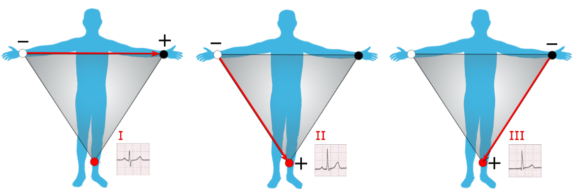
Photo used under Creative Commons from DJANDYW.COM & DJANDYW.TV AKA ANDREW WILLARD
